

- Home
- Our Services
- Ultrasound Imaging
Ultrasound Imaging

Gentle imaging with immediate clarity
Ultrasound Imaging
Ultrasound/Sonography is a method by which internal organs, -structures and blood flow are imaged in real-time, using high-frequency sound waves.
It is a safe, painless procedure as it does not utilise radiation (used for X-rays or CT scans).
Ultrasound is used in the evaluation of many different clinical conditions and can also be used to guide other procedures such as needle biopsies and interventional procedures.
Some ultrasound procedures may require special preparations.
When making your appointment our reception staff will inform you of these preparations, if applicable.

What to Expect
Your Visit Step by Step
You will be advised, when making your booking, of any preparation required before the scan. We suggest that you wear loose, comfortable clothing and preferably no jewellery.
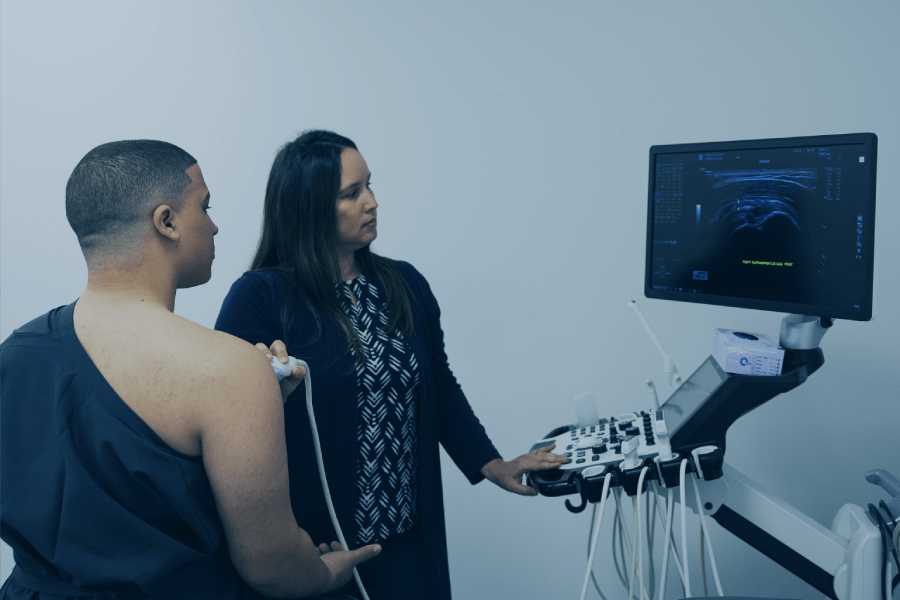
For certain examinations, you may be provided with, and asked to change into a gown prior to the examination. This serves simply to assist the sonographer in accessing the area to be scanned.
The examination will be performed by a qualified sonographer.
A water-based gel will be applied to the area to be scanned and a device, called a transducer, is used to acquire the images. This gel is easily wiped off and leaves no residue.
The ultrasound examination itself is painless. However, in the event that you have an injury or painful clinical condition, you may experience some discomfort as the examination requires the sonographer to apply a degree of pressure on the area being imaged, in order to adequately visualise the internal organs and structures.
The duration of the examination is dependent on the type of ultrasound performed. The average time for procedure: 20 – 40 minutes
The average time is dependant on the area of study, the complexity of case and number of images required.
Before and after the Ultrasound
Before the Ultrasound:
Ultrasound imaging for certain examinations is however optimized when the appropriate preparatory procedures have been followed. Therefore, in the event of a specialised examination, you will be advised in advance of the procedure and preparation required for this.
*Please refer to your specific ultrasound procedure for basic preparatory information.
For both general and specialised Ultrasound examinations, patients are advised to leave all jewellery at home and to wear loose, comfortable clothing.
After the Ultrasound:
Types of Ultrasounds
Abdominal Ultrasound:
Ultrasound is also used to guide interventional procedures such as liver and renal biopsies.
The preparation usually involves fasting for 6 hours and you may also be asked to drink water to fill the bladder before your examination. You may be asked to change into a gown before your examination.
Breast Ultrasound:
Breast ultrasound can be used as a primary examination in young patients, but is not recommended as a replacement for mammography in patients older than 40.
Ultrasound is also used to guide biopies or cyst aspirations, and for lesion localization prior to surgical excision.
Doppler Ultrasound:
No special preparation is necessary. The arterial Doppler examination is a longer procedure and, if not an emergency, booking an appointment is required.
Elastrography:
Your referring doctor may order elastography if you have risk factors, signs or symptoms of cirrhosis.
Elastography is a test that uses painless, low-frequency vibrations to check the elasticity of your body’s organs.
When tissues lack elasticity and are stiff, it may signal disease, especially in the liver.
What to expect:
Before your Elastography scan, our friendly reception team will inform you of any preparations prior to your scan.
On the day of the Elastography scan, the radiologist will apply a clear, water-based gel to your skin to help reduce friction.
While moving the handheld transducer, the wand will send painless, low-frequency vibration into your body.
The computer will turn these vibrations into an image of the relevant organ, which will help the radiologist determine the size, shape and consistency – including stiffness or wether it’s filled with fluid.
The whole procedure normally takes less than 15 minutes.
Your images will be shared directly with your referring doctor and shared with you via the MyMorton platform
MSK Ultrasound:
You may be asked to undress to expose the area that needs to be examined. You will be given a gown to wear, if necessary. No special preparation is needed.
Booking an appointment is required as some of the ultrasound examinations require specialised radiological skills.
Neonatal Ultrasound
Neonatal Brain Ultrasound:
While the anterior fontanelle of a newborn baby’s skull is open, ultrasound can be used, up to the age of six months, to examine the brain and the cerebral blood vessels. It is painless and requires no sedation or preparation.
Neonatal Hip ultrasound
Ultrasound of the hips is indicated for breech deliveries or if there is a clinical suspicion of hip dysphasia/instability. The examination is safe and painless. However, often babies will cry when being examined, particularly if they have to be held still in position. It usually is helpful to feed your baby just before the ultrasound study as baby will then be more relaxed.
Obstetric Ultrasound*
All other obstetric scans, including scans to determine fetal age, should be performed by a sub-specialist in obstetric ultrasound. Please consult your gynaecologist for a referral to the appropriate institution.
A full bladder is preferable.
Pelvic Ultrasound*
You will be asked to attend the examination with a full bladder to ensure optimal visualisation.
If your bladder is not sufficiently filled, you may be asked to drink water and wait until the bladder is full before rescanning.
Small parts Ultrasound*
For more information on any of the above specialised procedures, please contact enquiries@morton.co.za or contact any of our branches and chat to one of our receptionists who will be able to assist you with the necessary information.
Trans-vaginal Ultrasound:
You will be asked to empty the bladder, remove all clothing from the waist down, and be given a gown to wear.
The specially designed transducer, is covered with a sterile condom, lubricated with sterile gel and gently introduced into the vagina.
You may experience mild discomfort but this examination is usually very well tolerated.
The examination does not last longer than 5-10 minutes.
Trans-rectal Ultrasound*
This examination may be combined with a biopsy of the prostate, if indicated. The examination is generally less uncomfortable than a routine digital examination by your doctor.
The biopsy procedure requires several needle pricks through the wall of the rectum directly into the prostate gland and can take another five to ten minutes – it is a mildly uncomfortable procedure.
After the procedure, you can expect some rectal bleeding and there is usually blood staining of the urine for a few days.
This is a highly specialized examination and booking an appointment is essential.
Preparation requires an empty rectum, so a laxative suppository is inserted 2 hours before the procedure. If a biopsy of the prostate is planned, antibiotic tablets will be prescribed, to be taken 2 hours prior to the test.
A preparation pack with full instructions, tablets and suppository can be obtained from reception at any of our hospital branches.
We usually perform a routine abdominal scan first with a full bladder to assess the kidneys and the ability of the bladder to empty, so you will be asked to drink three glasses of water an hour before your appointment.