

MRI Scans and Magnetic Resonance Imaging

Deep Diagnostic Insight
MRI
Magnetic resonance imaging (MRI) uses a powerful magnetic field, radio waves and a computer to produce detailed images of the body’s internal structures that are clearer, more detailed and more likely to identify and accurately characterize disease than other imaging methods.
It is used to evaluate the body for a broad range of conditions, including cancer, heart- and vascular diseases and muscular- and bone abnormalities.
MRI is non-invasive and does not use ionizing radiation. If you have been referred by your physician to undergo an MRI at our Cape Town MRI or Richards Bay MRI centres, you can make your appointment via our centralized MRI Booking Centre.
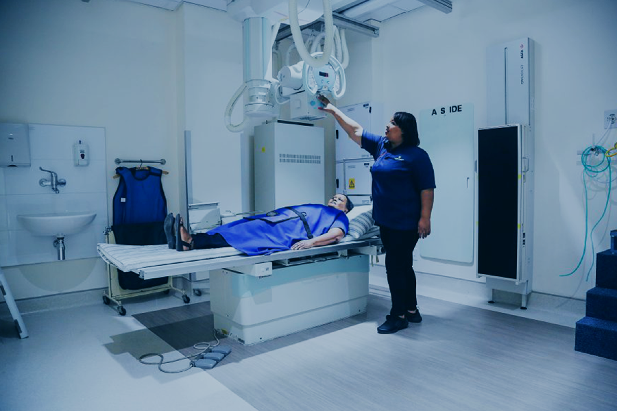
Referred by a Physician
If you have been referred by your physician to undergo an MRI at our Cape Town MRI or Richards Bay MRI centres, you can make your appointment via our centralized MRI Booking Centre.
You can contact them on:
(021) 276 1253
mribookings@morton.co.za
Please note: This number is only available for MRI bookings.
Our staff will inform you of specific preparations that might be required for your MRI procedure.
Tell our receptionist about any health problems (incl. allergies, implants, stimulators, etc.), recent surgeries or if you have an aversion to enclosed spaces.
Also, share with them if you suspect you might be pregnant.
We offer various types of MRI scans, including Brain MRI, MR Angiography, Prostate MRI, Breast MRI and MRI of the spine to name a few.

What to Expect
Your Visit Step by Step
At Morton & Partners, we try to make your experience with us as comfortable as possible; to this end many of our MRI machines use larger tunnels.
If you think that you might still feel anxious during the exam, you may ask our radiologist to administer a mild sedative prior to the exam.
Note that you would need to fast for at least 6 hours before your scan in order to take the sedative.
Most orthopaedic implants pose no risk, but you should always tell the radiographer, if you have any devices or metal in your body.
The magnetic field produced by our wide-bore MRI machine, is not harmful, but it may cause some medical devices (for example: pacemakers, aneurysm clips and prosthesis) to malfunction.
Our friendly booking receptionist will ask you a few protocol questions, when making your booking, in order to assign the right type of machine for your unique case.
On the day of the exam, you will also complete a patient questionnaire and consent form reconfirming the details you have submitted.
Before the exam starts you will be positioned inside the MRI unit. Devices that contain coils capable of sending and receiving radio waves may be placed around or adjacent to the area of the body being studied.
While you are in the machine, the radiographer will perform the examination while working outside of the room on a computer. You will be able to communicate with the radiographer for the duration of the exam.
An alert button is available if you feel too uncomfortable and would like to come out of the machine. If a child is being examined, a general anaesthetic may be required and a parent may be permitted to stay in the room.
The success of the MRI relies on your cooperation, since any movement will compromise the scan quality.
An injection of contrast material, to enhance the visibility of certain tissues or blood vessels, may be needed. This is generally administered more than half-way through the examination. To allay the prolonged noise during the examination, headphones are available with music of your choice.
Why are MRI rooms always cold?
The MRI room is kept cold (17 – 21° C ) to ensure an appropriate working environment for the machine’s magnets.
Under normal circumstance, our MRI radiographer will provide the patient with a blanket, however, following the recent outbreak of Covid-19 and the hygiene protocol associated with it, we will not be providing patients with blankets when undergoing a MRI scan at any of our departments. We, therefore, encourage patients to bring warmer clothes (for example cotton lounge wear or winter pajamas) for the exam. Depending on the type of MRI study you might be required to wear the provided medical gown.
Note the clothing should have no jewellery, zippers or wiring as this will affect the overall image quality.
The average time for procedure: 20 – 60 minutes
The average time is dependant on the area of study, the complexity of case and anaesthetic are required.
OUR MRI BOOKING SERVICE
Centralised MRI Bookings, Simplified
Book your MRI with confidence through our dedicated Centralised MRI Booking Centre. Our experienced team coordinates appointments across our MRI locations, making the process seamless, efficient, and convenient for both patients and referring healthcare professionals.
Before and after the MRI
Before the MRI:
You can contact them on:
(021 276 1253)
mribookings@morton.co.za
Please note: This number is only available for MRI bookings.
Our staff will inform you of specific preparations or lab work that might be required for your MRI procedure.
When making your appointment, tell our receptionists about:
• Any health problems (for example: pacemaker, aneurysm clip, Neuro-stimulators and Cochlear implants)
• Recent surgeries
• Allergies
• An aversion to enclosed spaces
To ensure you are relaxed and comfortable during the exam our radiologist will administer a mild sedative prior to the exam. Roughly 1 in 20 patients will require medication, so don’t feel disconcerted to ask. You will, however, be instructed not to eat or drink anything from midnight the night before the exam (or 6 hours before your procedure). If you disregard this directive, we would need to reschedule your exam.
• Any possibility that you might be pregnant.
MRI exams have been successfully performed on pregnant women without any ill effects on them or their unborn baby for more than 35 years. However, because the unborn baby will be in a strong magnetic field, we recommend that you should not have this exam in the first three to four months of pregnancy unless the potential benefit from the MRI exam is assumed to outweigh the potential risks. Pregnant women should also not receive injections of gadolinium contrast material except when absolutely necessary for medical treatment.
Dental fillings are usually not affected by the magnetic field, but may distort images of the facial area or brain. So the radiologist should be aware of them.
You will be asked to remove anything that might degrade the quality of the MRI images.
Please wear loose, comfortable clothing and leave your jewellery (for example: earrings and watches) at home.
You may be asked to wear a gown.
On the day of the exam, you will also complete a patient questionnaire and consent form reconfirming the details you have already submitted when making your booking.
Please note that certain MRI procedures require special preparations (for example: blood test). Our friendly booking staff will, however, inform you of the specific preparations for your procedure. If you would like to know more about your procedure, please visit the specific procedure page.
PLEASE NOTE:
The MRI room is kept cold (17 – 21° C ) to ensure an appropriate working environment for the machine’s magnets.
Under normal circumstance, our MRI radiographer will provide the patient with a blanket, however, following the recent outbreak of Covid-19 and the hygiene protocol associated with it, we will not be providing patients with blankets when undergoing a MRI scan at any of our departments. We, therefore, encourage patients to bring warmer clothes (for example cotton lounge wear or winter pajamas) for the exam. Depending on the type of MRI study you might be required to wear the provided medical gown.
Note the clothing should have no jewellery, zippers or wiring as this will affect the overall image quality.
After the MRI scan:
What you need to know about MRI
Understanding the Cost of an MRI:
Booking Your MRI: The First Step
Cost of MRI: Medical Aid Rates
Co-Payments and Medical Aid Plan Details
Pre-Authorisation: A Critical Step
Private-Paying Patients: Standard Private Rates
Non-South African Residents: Payment Requirements
Why Transparency Matters
Contact Us for More Information
MRI Brain
It can be used to diagnose and evaluate many conditions, of which a few are listed:
• Finding the cause of headaches
• Tumours of the brain
• Early diagnosis of a stroke
• Developmental abnormalities of the brain
• Vascular pathology
• Disorders of the inner ear
• Pathological conditions of the eyes
• Disease of the pituitary gland
You will be required to lie on your back during the examination.
A special frame-like piece of equipment called a coil, will be placed over your face. However, it will not touch your face. An eye mask is available should you prefer, find out more about what you can expect on our What to Expect MRI page.
Your head will be positioned in the middle of the MRI magnet, which means that only half of your body will be inside the “tunnel”.
The success of the MRI relies largely on your cooperation, since any yawning, coughing, chewing, snoring or excessive movement of the eyes or face will interfere with the scan.
This may cause the images to be less than optimal.
MR Breast
Before the Breast MRI scan:
You can contact them on:
(021 276 1253)
mribookings@morton.co.za
Please note: This number is only available for MRI bookings.
Our staff will inform you of specific preparations or lab work that might be required for your MRI procedure.
After the breast MRI:
Digital images and reports are available to your referring doctor as soon as the radiologists have viewed the examination and reported on the findings.
The radiologist, on occasion, may discuss preliminary results with you at the time of scanning, if necessary and appropriate
MR Arthrography
MR Arthrography:
Firstly, a thin needle is used to inject the contrast into the joint.
The addition of this fluid enhances the quality of the examination.
The radiologist may use a local anaesthetic to numb the injected area.
In some cases, a small amount of medication may also be administered.
During the injection of the contrast agent, patients may feel slight, temporary pressure or discomfort as the joint is distended.
Following the arthrography, the MRI scan is conducted for the defined area.
After the procedure:
There may be mild discomfort following the exam, but this should abate within 1-2 days.
You may apply ice to the joint and take a mild analgesic. Avoid aspirin.
MR Angiography
• Inflammation and aneurysms in the aorta or in other major blood vessels
• Atherosclerotic disease in the arteries of the neck, which may limit blood flow to the brain and cause a stroke
• Aneurysms or arteriovenous malformations in the brain
• An atherosclerotic disease that has narrowed the arteries to the legs
• To guide surgeons making repairs to diseased blood vessels, such as implanting or evaluating a stent
• Identify dissection or splitting in the aorta in the chest or abdomen or its major branches
The exact nature of the scan depends on the vessel that needs to be examined and the procedure varies accordingly.
MR Enterography (MRE)
MR Enterography (MRE):
It is non-invasive and does not use ionizing radiation.
Before the exam, oral and intravenous contrast material will be administered to highlight the small intestine. A drug may also be administered to decrease movement of the bowel, which can interfere with the images.
Special preparations for MRE:
You will be instructed to follow either a low faecal diet or be provided with a laxative (PicoPrep) which needs to be taken one day before the exam.
Our team might also ask you to arrive 1 hour before the appointment to drink the bowel prep.
When making your booking you will be provided with a Colon Preparation for Radiological examination document which outlines the specific instructions to be followed.
MRI Spine
It is typically used to demonstrate and evaluate spinal anatomy, anatomical variations and pathology such as:
• Bulging or degenerated discs causing back pain
• Compressed and inflamed spinal nerves
• Spinal infections
• Spinal tumours and / or metastases
• Spinal fractures and its impact on the spinal cord.
MRI for the Hepatobiliary and pancreatic/liver systems
Working in conjunction with leading hepatobiliary surgeons and physicians, our internationally trained hepatobiliary radiologist uses this MRI procedure to examine and diagnose conditions related to your digestive system (the liver, pancreas, bile ducts and the gallbladder).
Special preparation for Magnetic resonance cholangiopancreatography (MRCP)
Due to the nature of the area to be examined, you will be instructed to stop drinking and eating (Nil per mouth) 6-8 hours before the exam.
The appointment could take up to an hour to complete.
MRI Gynae Pelvis
Overview:
An MRI of a woman’s pelvis area will provide the referring practitioner with detailed images of the bones, muscles and organs in that region.
Organs found in the pelvis that are specific to women include the uterus, ovaries, fallopian tubes, cervix, vagina, rectum, bladder and urethra. For example, an MRI scan can help to evaluate the cause of pelvic pain in women or other anomalies in the abdominal organs.
For more information on the MRI of the pelvis.
There are a number of conditions that can arise in the pelvis area. For women especially, this is an important part of the body because it houses their reproductive organs.
MRI technology is non-invasive and enables healthcare practitioners to examine the inside of the human body in high detail.
MRI stands for Magnetic Resonance Imaging and its introduction represented a huge milestone for the medical world. An MRI uses a powerful magnetic field and radio waves to produce detailed images of the body’s internal structure. In comparison to other imaging methods, the images from an MRI are clearer, more detailed and more likely to identify and accurately evaluate the body for a broad range of conditions. An MRI does not use ionising radiation.
Morton & Partners are leaders in the radiology landscape and have more than 25 expert diagnostic radiologists in their service. They offer a broad range of sub-specialty expertise which is supported by their continued investment in imaging technology and services.
What to expect during an MRI Gynae Pelvis
An MRI is painless, and the patient is made to feel as comfortable as possible. If the patient feels though that they may feel anxious during the procedure, a mild sedative can be administered prior to the exam. Headphones are also available for the patient to listen to music during the exam.
The patient will be able to communicate with the radiographer at all times during the exam and an alert button is available if at any time the patient feels uncomfortable and would like to come out of the machine.
The magnetic field produced by the MRI machine is not harmful, but may cause some medical devices to malfunction (e.g. pacemakers, aneurysm clips and prosthesis). If the patient has any devices or metal in their body, please discuss these with our knowledgeable booking agent when making an appointment so the right type of machine can be assigned for the exam.
Devices that contain coils capable of sending and receiving radio waves may be placed around or adjacent to the area of the body being studied. The digital imaging is then fed from the MRI machine to a computer operated by the radiographer performing the examination outside of the exam room.
The patient can expect an MRI Gynae to be completed in 45 minutes or less. Digital images and reports will be made available to the referring physician once the radiologist has completed their diagnosis. Access to reports and images are stored safely online on the Morton & Partners website portal for the referring practitioner to access.
Making an appointment for an MRI Gynae
We have a centralised MRI booking centre for appointments at our Cape Town MRI or Richards Bay MRI centres.
The Morton & Partners MRI Booking Centre can be contacted on:
Tel: (021) 276 1253
Email: mribookings@morton.co.za
When contacting the booking centre, the patient will be advised of specific preparations that may be required for the MRI procedure. On booking, please share information on any health problems (including allergies, implants, stimulators, etc.), recent surgeries, and if the patient has an aversion to enclosed spaces.
The patient should wear loose, comfortable clothing and leave jewellery, including their watch and wedding ring, at home. The patient may be asked to wear a gown. If the patient has received a sedative, they will not be allowed (by law) to drive after the exam. It is required that someone drive the patient home.
The cost of an MRI Gynae Pelvis
We can provide assistance should your medical aid scheme require pre-authorisation before the procedure. Please note that pre-authorisation is not a guarantee of payment and the account remains the responsibility of the patient to settle.
For private-paying patients, the account will be rendered at the standard private rate of Morton & Partners.
Special preparation for an MRI Gynae
One of our expert consultants will advise you of any special preparations prior to the MRI Gynae.
MRI Prostate
Overview:
An MRI of the prostate is a very useful exam, especially when used in combination with the clinical history and patient information provided by the referring physician.
Prior to having an MRI of the prostate, patients can benefit from prostate screening tests, including a Digital Rectal Exam (DRE) or a Prostate-Specific Antigen (PSA) blood test.
Magnetic Resonance Imaging (MRI) uses a powerful magnetic field and radio waves to produce detailed images of the body’s internal structure.
In comparison to other imaging methods, the images from an MRI are clearer, more detailed, and more likely to identify and accurately evaluate the body for a broad range of conditions.
An MRI is also non-invasive and does not use ionising radiation.
An MRI of the prostate may be requested to help diagnose infection, benign prostatic hyperplasia (BPH), or congenital abnormalities.
The MRI scanner gives radiologists an excellent view of the prostate, allowing urologists to perform a targeted biopsy of the most suspicious region.
What to expect during an MRI of the prostate?
Devices that contain coils capable of sending and receiving radio waves may be placed around or adjacent to the area of the body being studied. An MRI of the prostate may also be performed using an endorectal coil – a thin wire covered with a latex balloon, that is inserted a short distance into the rectum.
The magnetic field produced by the wide-bore MRI machine is not harmful, but may cause some medical devices to malfunction (e.g. pacemakers, aneurysm clips and prosthesis). If the patient has any devices or metal in their body, please discuss these with our knowledgeable booking agent when making an appointment so the right type of machine can be assigned for the exam.
The digital imaging from the MRI machine is fed to a computer operated by the radiographer performing the examination outside of the exam room. The patient will be able to communicate with the radiographer at all times during the exam and an alert button is available if at any time the patient feels uncomfortable and would like to come out of the machine.
The patient can expect an MRI of the prostate to be completed in 45 minutes or less. Digital images and reports will be available to the referring physician once the radiologist has completed their diagnosis. Access to reports and images are st
How to make an appointment for an MRI of your prostate?
We have a centralised MRI booking centre for patients who need to make an appointment at one of our Cape Town MRI or Richards Bay MRI centres.
The Morton Partners MRI Booking Centre can be contacted on:
Tel: (021) 276 1253
Email: mribookings@morton.co.za
Our highly efficient, courteous and compassionate staff will guide you during the procedure to ensure your comfort, safety and privacy.
When contacting the booking centre, the patient will be advised of specific preparations that may be required for the MRI procedure. On booking, please share information on any health problems (including allergies, implants, stimulators, etc.), recent surgeries, and if the patient has an aversion to enclosed spaces.
The patient should wear loose, comfortable clothing and leave jewellery such as their watch and wedding ring at home. The patient may be asked to wear a gown.
If the patient has received a sedative, they will not be allowed (by law) to drive after the exam. It is required that someone drive the patient home.
What is the cost of an MRI of the prostate?
Should pre-authorisation be required by the medical aid scheme, we can provide assistance in applying for pre-authorisation.
Please note that pre-authorisation is not a guarantee of payment and the account remains the responsibility of the patient to settle.
For private-paying patients, the account will be rendered at the standard private rate of Morton & Partners.
All non-South African residents will be required to settle their radiology account in full on the day of examination or discharge from the hospital if admitted as an in-patient at the time of the examination.
Special preparation for an MRI Prostate:
Lenolax (a laxative enema) must be taken an hour prior to the procedure. Please consult with our expert booking agents when making the appointment.
Note: Lenolax is available without a prescription from your local pharmacy. It is also available at any of the Morton & Partners MRI branches
MRI Pelvis
Overview:
A man’s pelvis has structural differences to a female’s pelvis. For one, a man’s pelvic bones are smaller and narrower to a woman’s pelvic bones which will also support her reproductive organs. To read more on what can be detected on an MRI of a woman’s pelvis, visit our page at MRI Gynae.
An MRI of the pelvis creates medical imaging of the bones (skeletal system), organs (endocrine system), tissue, blood vessels (circulatory system) and muscular system in the region. The pelvic girdle forms part of the body’s skeletal system and is made up of three bones: ilium, ischium and pubis. Organs in that region of the male anatomy include the bladder, prostate, uretha and rectum.
The urinary bladder is a hollow muscular organ that is also examined by an MRI of the pelvis. The pelvic floor is the set of muscles that support the pelvic organs of the bladder and bowel.
For information on the MRI of the prostate.
An MRI, also known as Magnetic Resonance Imaging, uses a powerful magnetic field and radio waves to produce detailed images of the body’s internal structure. In comparison to other imaging methods, the images from an MRI are clearer, more detailed and more likely to identify and accurately evaluate the body for a broad range of conditions. An MRI does not use ionising radiation.
What to expect during an MRI of the pelvis
An MRI is painless, and the patient is made to feel as comfortable as possible. If the patient feels though that they may feel anxious during the procedure, a mild sedative can be administered prior to the exam. Headphones are also available for the patient to listen to music during the exam.
The patient will be able to communicate with the radiographer at all times during the exam and an alert button is available if at any time the patient feels uncomfortable and would like to come out of the machine.
The magnetic field produced by the MRI machine is not harmful, but may cause some medical devices to malfunction (e.g. pacemakers, aneurysm clips and prosthesis). If the patient has any devices or metal in their body, please discuss these with our knowledgeable booking agent when making an appointment so the right type of machine can be assigned for the exam.
Devices that contain coils capable of sending and receiving radio waves may be placed around or adjacent to the area of the body being studied. The digital imaging is then fed from the MRI machine to a computer operated by the radiographer performing the examination outside of the exam room.
The patient can expect an MRI of the pelvis to be completed in 45 minutes or less. Digital images and reports will be made available to the referring physician once the radiologist has completed their diagnosis. Access to reports and images are stored safely online on the Morton & Partners website portal for the referring practitioner to access.
Making an appointment for an MRI of the pelvis
We have a centralised MRI booking centre for appointments at our Cape Town MRI or Richards Bay MRI centres.
The Morton & Partners MRI Booking Centre can be contacted on:
Tel: (021) 276 1253
Email: mribookings@morton.co.za
When contacting the booking centre, the patient will be advised of specific preparations that may be required for the MRI procedure. On booking, please share information on any health problems (including allergies, implants, stimulators, etc.), recent surgeries, and if the patient has an aversion to enclosed spaces.
The patient should wear loose, comfortable clothing and leave jewellery, including their watch and wedding ring, at home. The patient may be asked to wear a gown.
If the patient has received a sedative, they will not be allowed (by law) to drive after the exam. It is required that someone drive the patient home.
The cost of an MRI of the pelvis
We can provide assistance should your medical aid scheme require pre-authorisation before the procedure. Please note that pre-authorisation is not a guarantee of payment and the account remains the responsibility of the patient to settle.
For private-paying patients, the account will be rendered at the standard private rate of Morton & Partners.
Special preparation for an MRI of the pelvis
For example, the patient may be restricted on when to stop eating or drinking before the procedure. A low faecal diet may need to be followed, and a colon preparation may be required before the exam.
Note: Lenolax (a laxative enema) is available without a prescription from your local pharmacy. It is also available at any of the Morton & Partners MRI branches.