Women’s Imaging

Dedicated Women’s Care

Women’s Imaging

Morton & Partners is committed to providing a dedicated diagnostic and screening service in the field of women’s health and wellness.

From Mammography screening and breast MRI’s to Bone Mineral Density scans, our staff have been specially trained to provide these services in a relaxing, professional environment.

Early detection, expert assessment, and ongoing reassurance

Focused Care for Women’s Health

Mammography overview 

Mammography uses a low-dose X-ray system and high-contrast, high-resolution film for examination of the breasts. It plays a central part in early detection of breast cancers and can detect 85 to 90 percent of breast cancers in women. Breast Ultrasound Ultrasound of the breast is generally used in conjunction with mammography: in the presence of dense breast tissue, to further assess a palpable lump, or for an abnormality detected on mammography. It is a painless and safe examination. Breast ultrasound can be used as a primary examination in young patients, but is not recommended as a replacement for mammography in patients older than 40. Ultrasound is also used to guide biopies or cyst aspirations, and for lesion localization prior to surgical excision.

MRI of the breast overview

Magnetic resonance imaging (MRI) of the breast uses a powerful magnetic field, radio waves and a computer to produce detailed pictures of the structures within the breast. It is primarily used as a supplemental tool to breast screening with mammography or ultrasound. It may be used to screen women at high risk for breast cancer, evaluate the extent of cancer following diagnosis, or further evaluate abnormalities seen on mammography.

Breast MRI does not use ionizing radiation, and it is the best method for determining whether silicone breast implants have ruptured.

Bone Mineral Density Scan (BMD) overview:

BMD scans are simple, safe, and painless. Bone Mineral Density scans are extremely precise and are one of the most accurate methods available for measuring bone density. Using ultra-low dose x-ray, the BMD machine scans and measures bone mineral density at various parts of the body, usually the lumbar spine and hips. This highly sensitive test enables us to identify bone loss early and determine whether you are at risk for fracture, by comparing your bone density to your own age group and to a younger age group. The higher the density, generally, the lower the risk of fracture.

We can also monitor your response to treatments for osteoporosis by checking your bone density regularly. Please ensure that follow up BMD’s are scanned at the same facility as comparisons can only be done on the unit your previous scan was performed. Routine evaluations every two years may be needed to see a significant change in bone mineral density, decrease or increase. Few patients, such as patients on high dose steroid medication, may need a follow-up at six months.

Specialist care you can trust

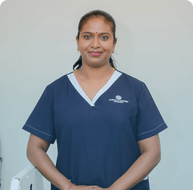
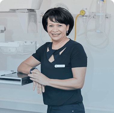
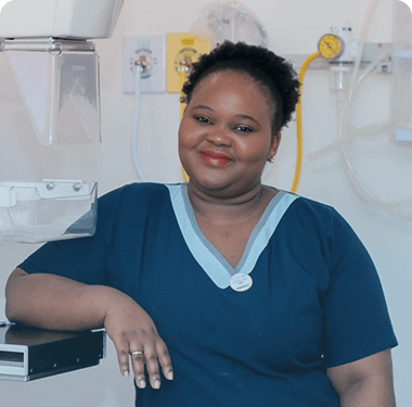
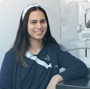
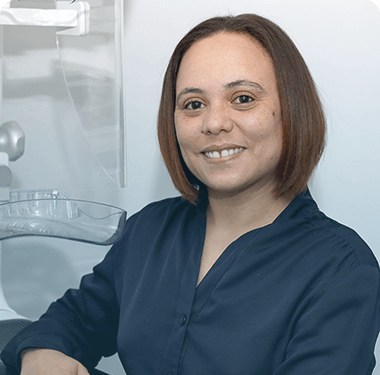
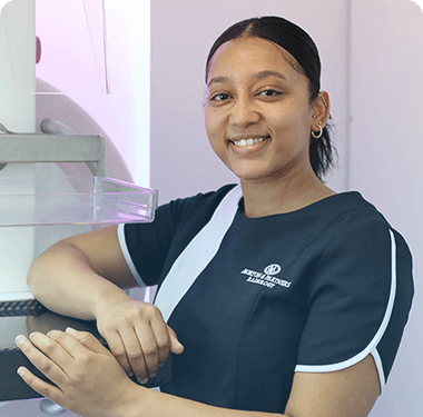
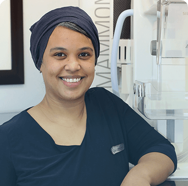
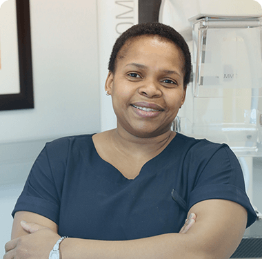
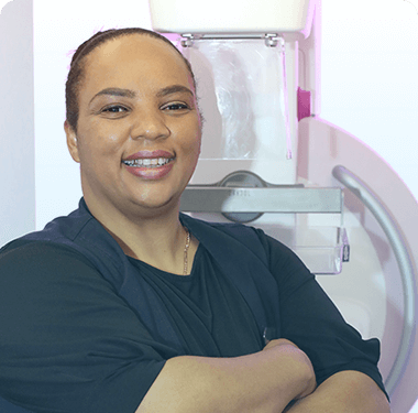
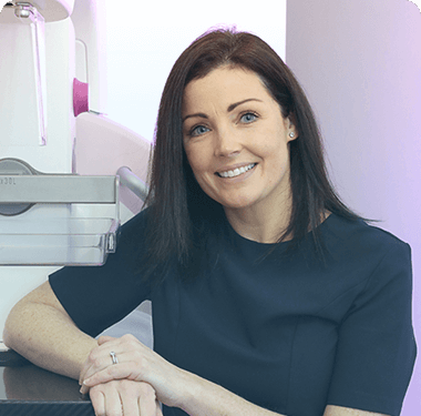
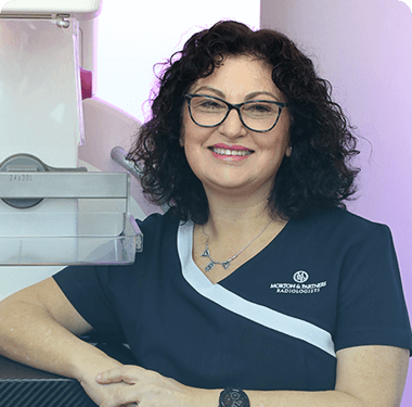
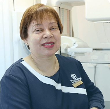
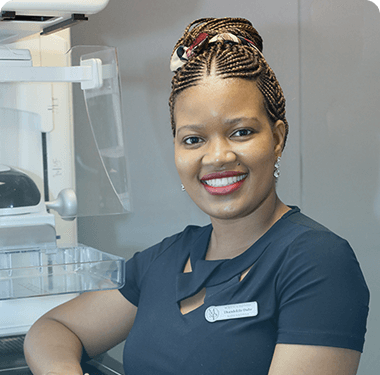
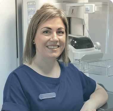
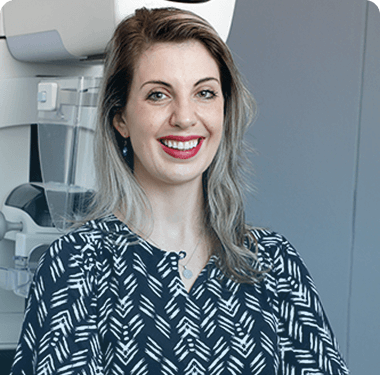
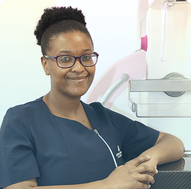
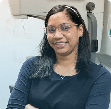
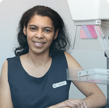
Meet your Mammographer

Our management team is comprised of experts from diverse industries. Supported and guided by the clinical team and Managing Partner, the team aims to drive strategic business objectives and innovation, whilst enriching our patient-centric culture.

Digital Mammogram booking request

    What to expect when going for a mammogram?

    What to expect when going for a mammogram:
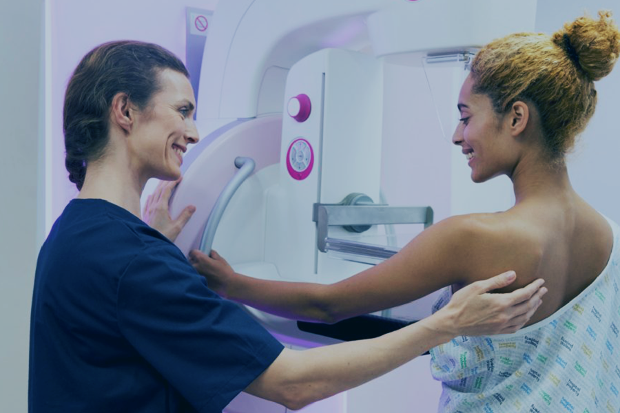
    During a mammogram, the radiographer will position you and image the breast. The breast is first placed on a special cassette and compressed with a paddle (made of clear Plexiglas or other soft plastic).

    Breast compression is necessary in order to
    • Even out the breast thickness so that all of the tissue can be visualized
    • Spread out the tissue so that small abnormalities aren’t obscured by overlying breast tissue
    • Allow the use of a lower x-ray dose since a thinner amount of breast tissue is being imaged
    • Hold the breast still in order to eliminate blurring of the image
    • Reduce x-ray scatter which also leads to poor image quality

    The radiographer will make the first x-ray exposure.

    You will be asked to change positions slightly between images.

    The process will then be repeated for the other breast.

    Generally, this is a painless procedure.

    You will feel pressure on the breast as it is squeezed by the compressor.

    Some women with sensitive breasts may experience discomfort.

    Be sure to inform the radiographer if pain occurs as compression is increased.

    The examination process may take up to half an hour.

    Sometimes additional images need to be taken from different angles or with differing degrees of compression to show internal breast structures more clearly.

    It may also be necessary to perform an ultrasound or MRI examination to add more information, particularly if breast tissue is dense, or if a lump can be felt or seen.

    What to expect when going for a Breast MRI?
    Please visit our MRI service page for a comprehensive review of what to expect and how to prepare for your breast MRI. Most MRI exams are painless, however, you might find it uncomfortable to remain still during the imaging process.

    For an MRI of the breast, you will lie face down on a platform specially designed for the procedure. The platform has openings to accommodate your breasts and allow them to be imaged without compression.

    The electronics needed to capture the MRI image are actually built into the platform. It is important to remain very still throughout the exam.
    Be sure to let the radiographer know if something is uncomfortable since discomfort increases the chance that you will feel the need to move during the exam.
    What to expect when going for a Bone Mineral Density (BMD) scan?
    Our Bone Mineral Density scan is a quick and painless procedure.

    You will be asked to lie down on a padded table.

    As a standard, your spine and one/both hips will be examined.

    In some cases, the forearm might also be scanned, if either the hip or spine is unavailable (usually due to surgery or implants).

    As any condition affecting bone density tends to affect the whole skeleton, a snapshot of a few sites is sufficient to establish the overall bone density.

    The BMD at the hip and spine has been shown to be the best way of predicting the risk of fracture.

    The BMD scan is a painless procedure.

    Before and after the Mammogram

    The body, especially the breast, goes through natural cycles, but being able to detect any abnormalities early will help increase your chances of overcoming any serious health risks.

    Mammograms play a crucial role in the detection of abnormalities in the breast and The American Cancer Screening guidelines recommend women between the ages of 40 to 44 should have the choice to start annual breast cancer screening with mammograms.

    Women, aged 45 to 54, should get mammograms every year, thereafter, women 55 and older should switch to mammograms every 2 years, or can continue their annual screening routine.

    Before the Mammogram:
    If you have been referred by your physician or would like to go for your routine check-up, you can book an appointment at any of our branches.

    • It is advised not to schedule your mammogram for the week before your menstrual cycle if your breasts are usually tender during this time. The best time to book an appointment is normally one week following your monthly cycle.
    • Always inform your doctor or radiographer if there is any possibility that you might be pregnant. Do not wear deodorant, talcum powder or lotion under your arms on the day of the exam as these can appear on the x-ray film as white spots mimicking pathology.
    • Before your mammogram, your mammographer will explain the procedure to you and complete a quick questionnaire with you to provide clinical history for the radiologist.
    • The mammogram is mostly pain-free depending on the machine and the density of your breast.

    Very important: Remember to make your previous mammogram images available to the radiologist at the time of the current examination.
    How is a mammogram performed:
    During a mammogram, the radiographer will position you and image the breast. The breast is first placed on a special cassette and compressed with a paddle (made of clear Plexiglass or other soft plastic).

    You will be asked to change positions slightly between images, and then the process will be repeated for the other breast.
    After the mammogram:
    Digital images and reports are available to your referring doctor

    Before and after the Breast MRI

    Before the Breast MRI scan:
    You can book your appointment via our centralised MRI Booking Centre, after being referred by your specialist/physician.

    You can contact them on:
    (021 276 1253)
    mribookings@morton.co.za

    Please note: This number is only available for MRI bookings.

    Our staff will inform you of specific preparations or lab work that might be required for your MRI procedure.
    After the breast MRI:
    If you have not been sedated, no recovery period is necessary. You may resume your usual activities and normal diet immediately after the exam.

    Digital images and reports are available to your referring doctor as soon as the radiologists have viewed the examination and reported on the findings.

    The radiologist, on occasion, may discuss preliminary results with you at the time of scanning, if necessary and appropriate.

    Before and after the Bone Mineral Density (BMD) scan

    BMD’s or Bone Density Scans are simple, safe, and painless. Bone Mineral Density scans are extremely precise and are one of the most accurate methods available for measuring bone density.

    Using ultra-low dose x-ray, the BMD machine scans and measures bone mineral density at various parts of the body, usually the lumbar spine and hips.

    Before the Bone Mineral Density scan:
    If you have been referred by your physician or would like to go for your routine check-up, you can book an appointment at any of our branches.

    • No special preparation is required for a BMD.

    It is helpful, but not essential, to wear loose fitting, comfortable clothing without metal buttons, buckles, fasteners or zippers, as metal objects interfere with the scan.

    A gown or sheet will be provided if clothing needs to be removed.

    A BMD scan does involve a very small dose of radiation, which makes this test unsuitable for women who are or might be pregnant.

    If you have had spinal surgery, particularly with metallic implants, or hip surgery (hip replacements, screws or pins), you will need to inform the radiographer performing the scan.

    They might decide to avoid that area.
    After the Bone Mineral Density scan:
    Digital images and reports will be made available to your referring doctor once our expert radiologists have completed their diagnosis.
    There are no side effects after the BMD scan.
    The Importance of Early Diagnosis
    Through early detection patients with osteopaenia (low bone mass) or osteoporosis can take action to stop the progressive loss of bone mass.

    By making positive lifestyle changes and by following appropriate treatment strategies in consultation with a doctor, osteoporosis can be prevented and treated.