

- Home
- Our Services
- GIT imaging
Barium Swallow and Fluoroscopy Imaging

Precise Functional Imaging
GIT Imaging
Gastro-Intestinal Imaging (a form of Fluoroscopy), uses contrast media and X-rays to examine the anatomy and its function.
As most Gastro Intestinal Imaging (GIT) studies require some form of preparation an appointment is essential – unless it is an emergency. The radiographer assisting the radiologist will help you at all times to ensure that you are as comfortable as possible during the examination.
The contrast medium generally used during the procedure is a barium-based preparation, but iodine preparations are also used in certain instances.
After you have been referred by your physician, you can book your appointment at any of our branches.
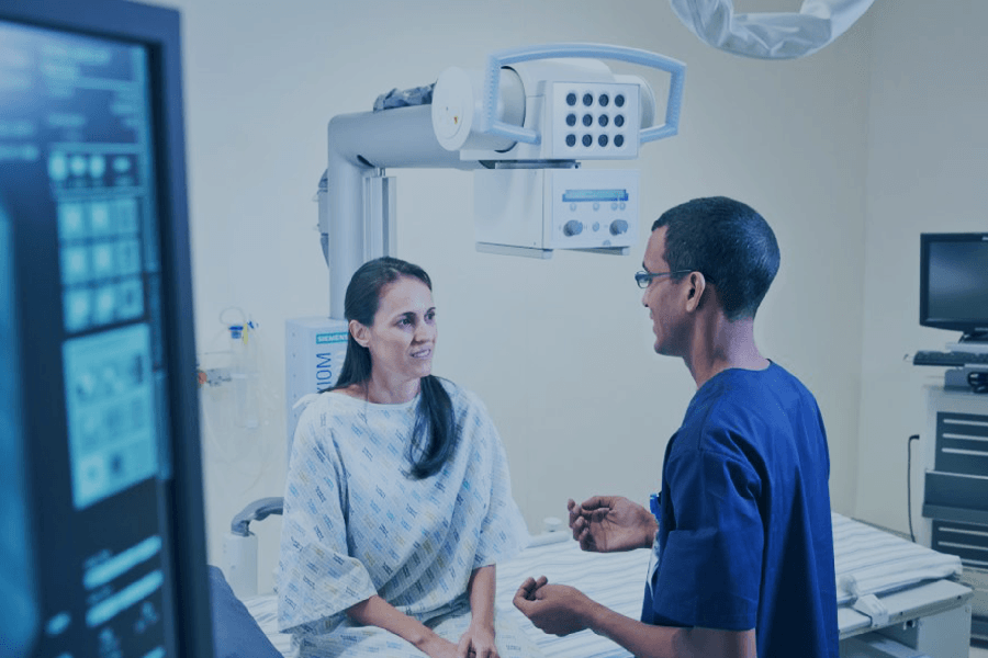

What to Expect
Your Visit Step by Step
As most Gastro Intestinal Imaging (GIT) studies require some form of preparation, an appointment is essential – unless it is an emergency.
When making your appointment our friendly receptionist will instruct you of any specific preparations required.
It is important that your colon be completely empty for the procedure, for that reason you will likely be asked not to eat 6 hours prior to your examination. And might be instructed to take a laxative (in either pill or liquid form) and/or to use an over-the-counter enema preparation the night before the examination and possibly a few hours before the procedure.
Please tell our staff when making the booking of any chronic medication you are taking or if you suspect that you might be pregnant. Many imaging tests are not performed during pregnancy so as not to expose the fetus to radiation. If an X-ray is necessary, precautions will be taken to minimize radiation exposure to the baby.
You will be required to undress and wear the gown provided.
The contrast administration is dependent on the area or organ that is to be examined (i.e. esophagus, stomach, small bowel or large bowel).
During the examination, you will be asked to follow specific instructions (depending on your examination type) given by the radiographer and to position yourself in order to best study the required part of the body.
The exam table will be positioned at different angles and your abdomen may be compressed to help spread the contrast material. Once the upper or lower (dependant on the area of study) GI tract is adequately coated with barium, still X-ray images will be taken and stored for further review.
Although the actual Gastro Intestinal Imaging (GIT imaging) is usually not painful, it may cause slight/moderate discomfort due to the contrast medium being swallowed.
Before and after GIT Imaging
Before GIT Imaging:
If you suspect you might be pregnant, notify our receptionist when scheduling the appointment, as additional precautions may need to be taken.
You will be requested not to eat or drink anything for 6 hours before your examination.
You may also be instructed to take a laxative (in either pill or liquid form) and/or use an over-the-counter enema preparation the night before the examination.
Possibly, this may need to be repeated a few hours before the procedure.
You will also be asked to wear a gown for the procedure.
The contrast will be administered before the imaging procedure starts.
After the GIT Test:
After the examination, you can resume a regular diet and take orally administered medications unless instructed otherwise by your doctor.
The contrast may colour your stool grey or white for 48 to 72 hours after the procedure. Sometimes the barium can cause temporary constipation, which is usually treated with an over-the-counter laxative. Drinking large quantities of fluids for several days following the test can also help.
If you are unable to have a bowel movement or if your bowel habits undergo any significant changes following the exam, you should contact your physician.
What you need to know about GIT Imaging
Barium Swallow (Gastrografin)
A series of x-ray images are taken while the patient swallows the contrast.
At the conclusion of the examination, a mild purgative is administered to facilitate the passage of the barium and the patient is encouraged to drink plenty of fluid.
Barium Meal (Gastrografin)
It can be performed using a single or double contrast technique where barium is swallowed and a series of X-rays is taken. The optimum way of performing this examination utilises the double contrast method which produces good distension as well as a good coating of the mucosal surface permitting demonstration of superficial abnormalities as well as larger lesions.
The examination commences with an intravenous injection of Buscopan (or equivalent) which causes relaxation of the smooth muscle in the stomach for 10 to 15 minutes.
This allows the stomach to be adequately distended and stops peristalsis for the period of the test.
A granular effervescent agent is then swallowed with a small amount of water.
This generates gas in the stomach to distend it. Thereafter you will be asked to swallow a small cup of barium while X-rays of the oesophagus are taken.
A further series of X-rays is taken showing these areas in a variety of projections to demonstrate any abnormalities.
The conclusion of the examination, a mild purgative is administered to facilitate the passage of the barium and the patient is encouraged to drink plenty of fluid.
The barium may colour your stools grey or white for 48 to 72 hours after the procedure. Sometimes the barium can cause temporary constipation, which is usually treated with an over-the-counter laxative. Drinking large quantities of fluids for several days following the test can also help.
If you are unable to have a bowel movement or if your bowel habits undergo any significant changes following the exam, you should contact your physician.
Barium Enema
Our expert radiographers will be there to guide you through the procedure.
You will be positioned on the examination table and an x-ray film is taken to ensure the bowel is clean.
After performing a rectal examination, the radiologist will then insert a small tube into the rectum and begin to instill, using gravity, a mixture of barium and water into the colon. Air may also be injected through the tube to help the barium thoroughly coat the lining of the colon. In some circumstances, the radiologist or referring physician may prefer a water and iodine solution rather than barium.
As the barium fills your colon, you will feel the need to move your bowel. You may feel abdominal pressure or even minor cramping. Most people tolerate the mild discomfort easily. The tip of the enema tube is specially designed to help you hold in the barium. If you are having trouble, let our team know.
Next, a series of x-ray images are taken. To improve the quality fo the image you must try to remain still during the examination. The radiographer will go into the adjoining room to activate the machine and will be able to see, hear and speak to you for the duration of the exam.
You may be repositioned frequently in order to image the colon from several angles. Some x-ray equipment will allow patients to remain in the same position throughout the examination.
When the examination is complete, you may be asked to wait until the radiologist determines that all the necessary images have been obtained.
Once the x-ray images are completed, most of the barium will be emptied through the tube. You will then be allowed to expel the remaining barium and air in the restroom.
A barium enema is usually completed within 30 to 60 minutes.