

- Home
- Our Services
- CT Imaging
Computed Tomography Imaging

High-Detail Imaging
CT Imaging
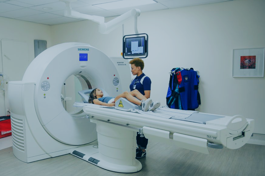
Computed Tomography, more commonly known as CT Imaging or CAT scan. The procedure uses special image data acquired at different angles whilst you, the patient, travel through the CT scanner on a bed and thereafter, advanced computer processing allows the radiologist to see cross-sectional images of the body.
CT imaging is one of the best tools for studying the head, neck, chest, abdomen and pelvis as well as complex fractures. It can also be used to guide biopsies, joint injections and other minimally invasive procedures.

What to Expect
Your Visit Step by Step
CT scanning causes no pain and with MDCT (Multidetector CT) the need to lie still for any length of time is reduced. Preparation for each body examination area will be different.
When making your booking at any of our branches, our friendly staff will inform you of how and what you need to do in order to prepare for your examination.
In some instances, you may be asked to swallow a liquid contrast material to help see the stomach, small bowel and colon better. Some patients find the taste mildly unpleasant, but most can easily tolerate it.
Commonly, a contrast material may be injected into a vein to define the blood vessels and to accentuate the difference between normal and abnormal tissue in organs like the liver, pancreas and kidneys.
A flush of heat together with a metallic taste at the back of the mouth are experienced but these usually disappear within a minute or two.
The total length of a CT scan procedure could take up to an hour. The actual scanning itself takes between a few seconds and a few minutes, depending upon the examination. Most of the procedure time is in preparation and confirmation that the images are of sufficient quality. In the case of children, a parent/carer may be allowed in the room to alleviate anxiety but will be required to wear a lead apron to prevent radiation exposure. You will be alone in the examination room for the duration of the scan. The radiographer is in an adjoining room and so will be able to see, hear and speak to you at all times. The average time for procedure: 20 – 60 minutes
The average time is dependant on the area of study, the complexity of the case and if any contrasting agents need to be administered.
Before and after the CT
Before the CT scan:
Should you have allergies, asthma, heart conditions, renal disease or diabetes mellitus (particularly important for patients taking Metformin) please bring this to the attention of the radiologist as such conditions may need special precautions to be implemented.
For this purpose, except in emergencies, booking an appointment is essential.
Clear instructions of special preparations will be given at the time of booking your appointment
If you suspect you might be pregnant notify our receptionist in advance, as this type of examination may be supplanted by an alternative procedure.
The radiographer begins the procedure by positioning you on the CT table. If necessary, pillows will be used to help hold you in the correct position during the scan.
As the study proceeds, the table will move through the CT scanner while the images are taken.
The amount of movement is dependent on the area of the body being examined. If a larger area is scanned you will definitely feel the table moving.
Sometimes you will be asked to hold your breath during the scan.
After the procedure:
What you need to know about CT Imaging
Cost of CT
We advise patients to review their medical aid plan details prior to making their appointment. Immediate settlement of the shortfall will be required at the time of service.
Should pre-authorisation be required by the medical aid scheme, we can provide assistance in applying for pre-authorisation.
Please note that pre-authorisation is not a guarantee of payment and the account remains the responsibility of the patient to settle.
For private-paying patients, the account will be rendered at the standard private rate of Morton & Partners.
All non-South African residents will be required to settle their radiology account in full on the day of examination or discharge from the hospital if admitted as an in-patient at the time of the examination.
Advanced 3D Imaging
Cone Beam CT (CBCT)
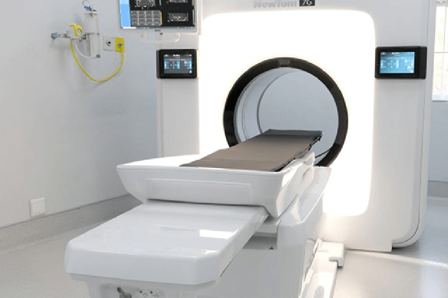
The NewTom 7G Cone Beam CT (CBCT) is a cutting-edge technology revolutionizing radiology and diagnostic imaging. This advanced scanner offers a range of benefits and applications, ensuring precise and detailed imaging for various clinical needs.
Cone Beam CT (CBCT)
Benefits of the CBCT scanner:
Versatile Imaging:
For the first time, Cone Beam technology is applicable to all body areas, including the spine, shoulders, and hips. This capability is particularly useful in reducing artefact from metalwork in patients with instrumentation which has historically been difficult to image in conventional CT.
High Resolution:
With a resolution of up to 90 µm, the CBCT can analyse small and complex structures, such as the inner ear ossicles, with exceptional precision and detail.
Suitable for All Patients:
The NewTom 7G is designed to accommodate patients of all ages and sizes, including pediatric and heavier individuals up to 215 kg. Its automated function adjusts imaging settings based on the patient’s build, ensuring safety and precision.
Customized Imaging:
An automated function adjusts the field of view and X-ray dose based on the patient’s build, making it ideal for pediatric patients. Additionally, options including the Ultra Rapid scan mode or ECO Low Dose scan can reduce X-ray exposure. Pulsed emission CBCT technology enables X-ray imaging, further minimizing exposure.z
Expanded Procedure Capabilities:
In addition to diagnostic imaging, the NewTom 7G enables a wide range of minor procedures, including CT guided joint infiltrations, arthrograms, lumbar punctures (LP), nerve blocks, and pain management interventions. This versatility enhances the utility of the CBCT system, providing comprehensive care options for patients in need of both imaging and minimally invasive procedures with reduced radiation exposure and exceptional accuracy.
The NewTom 7G Cone Beam CT Clinical Applications
Dental Implant Planning:
Provides precise assessment of bone quality, density, and anatomical structures before dental implant placement.
Orthodontic Evaluation:
Assists in orthodontic treatment planning, root position assessment, and identification of anatomical irregularities.
Maxillofacial Surgery:
Crucial for surgical planning, assessing facial fractures, and diagnosing temporomandibular joint (TMJ) conditions. The NewTom 7G also aids in post-treatment evaluation, offering detailed reviews of surgical hardware positioning to optimize patient recovery and outcomes following maxillofacial procedures.
Ear, Nose, and Throat (ENT) Conditions:
Ideal for diagnosing sinus issues, evaluating middle and inner ear pathologies, and assessing complex head and neck disorders.
Orthopedic Assessment:
Beneficial for evaluating small joints, bones, and soft tissues in orthopedic cases, including pre-operative planning and fracture evaluations. CBCT is particularly useful in reducing artefact from metalwork in patients with instrumentation which has historically been difficult to image in conventional C
CT Abdomen & Pelvis
CT Abdomen & Pelvis
The CT Abdomen is useful in diagnosing the cause of abdominal/pelvic pain and disease of the internal organs, small intestine and colon which can be caused by the following:
• Infections such as pyelonephritis or abscesses.
• Inflammatory conditions – appendicitis, diverticulitis, pancreatitis, Crohn’s disease.
• Renal (kidney) stones.
• Cancers of the stomach, colon, liver, kidneys, bladder, pancreas as well as lymphoma.
• Injuries to the internal organs.
• Abdominal aortic aneurysms (see CT Angiogram).
• Pre- and post-surgical assessment.
• Guidance for biopsy/drainage of pathological findings.
Special preparation for CT Abdomen & Pelvis:
You may have to drink a special fluid, which will fill the bowel and make it easier to recognize pathology around the colon and small intestines.
If you have had a barium examination recently please notify reception.
Angiography
The examination requires a high-level of skill and our staff undergoes special training to be able to scan the heart and process the images to create angiographic and 3D images of the heart and coronary vessels.
When you arrive your heart rate and blood pressure will be monitored. If your heart rate is above 80bpm a beta-blocker tablet will be given, after consultation with the radiologist or cardiologist. The heart rate is monitored again 45 minutes later and if it has slowed sufficiently the radiographer will start the scan. If your heart rate is still not optimal, you may be given another tablet and will be asked to wait another 30 minutes.
Your co-operation is critical to ensure the success of the scan. The radiographer will give you special breathing instructions and a breathing exercise will be performed a number of times to monitor the heart rate during a breath hold. An intravenous line will be inserted for the contrast injection. A vasodilator will be sprayed under the tongue just before the scan to dilate the cardiac vessels for the scan. This may cause a slight headache, which resolves rapidly.
Two scans will be done. The first will identify any calcified plaque in the coronary arteries. The number and size of these plaques are used to calculate the Agatston Calcium score, which quantifies the relative risk for cardiac disease.
The second scan uses the intravenous contrast agent and utilizes special technology that enables the scanner to acquire images at the same instant in every heartbeat, creating an angiographic image without movement. This is very useful to identify and diagnose the following conditions:
• Abnormal coronary artery origins or courses
• Narrowing/occlusion of the coronary arteries
• Patency of arteries after coronary stenting
• Electro-Anatomical Mapping of the left atrium prior to ablation therapy
If significant pathology is identified you may be referred for a conventional cardiac angiogram. The Cardiac CT Angiogram, however, remains a non-invasive, low-risk first investigation in patients at risk for cardiac disease.
The total time spent in our department can be 1 – 2 hours.
After the scan, the post-processing is done with great care to ensure accuracy and the radiologist carefully reviews the images. The images and a detailed report by our specially trained radiologist will be sent to your referring doctor.
Special preparation for a CT of the Coronary Arteries:
This examination is highly specialized and relies on your co-operation. Please ensure that you advise the receptionist and radiographer of the following conditions at the time of booking the appointment so that precautions can be taken:
• Asthma
• Diabetes
• Renal disease
• Allergies
• Pregnancy
• Heart disease
You will be asked to avoid intense activity, any caffeine and not smoke on the day of the scan.
CT Angio of the Coronary Arteries
CT Angio of the Coronary Arteries:
The examination requires a high-level of skill and our staff undergoes special training to be able to scan the heart and process the images to create angiographic and 3D images of the heart and coronary vessels.
When you arrive your heart rate and blood pressure will be monitored. If your heart rate is above 80bpm a beta-blocker tablet will be given, after consultation with the radiologist or cardiologist. The heart rate is monitored again 45 minutes later and if it has slowed sufficiently the radiographer will start the scan. If your heart rate is still not optimal, you may be given another tablet and will be asked to wait another 30 minutes.
Your co-operation is critical to ensure the success of the scan. The radiographer will give you special breathing instructions and a breathing exercise will be performed a number of times to monitor the heart rate during a breath hold. An intravenous line will be inserted for the contrast injection. A vasodilator will be sprayed under the tongue just before the scan to dilate the cardiac vessels for the scan. This may cause a slight headache, which resolves rapidly.
Two scans will be done. The first will identify any calcified plaque in the coronary arteries. The number and size of these plaques are used to calculate the Agatston Calcium score, which quantifies the relative risk for cardiac disease.
The second scan uses the intravenous contrast agent and utilizes special technology that enables the scanner to acquire images at the same instant in every heartbeat, creating an angiographic image without movement. This is very useful to identify and diagnose the following conditions:
• Abnormal coronary artery origins or courses
• Narrowing/occlusion of the coronary arteries
• Patency of arteries after coronary stenting
• Electro-Anatomical Mapping of the left atrium prior to ablation therapy
If significant pathology is identified you may be referred for a conventional cardiac angiogram. The Cardiac CT Angiogram, however, remains a non-invasive, low-risk first investigation in patients at risk for cardiac disease.
The total time spent in our department can be 1 – 2 hours.
After the scan, the post-processing is done with great care to ensure accuracy and the radiologist carefully reviews the images. The images and a detailed report by our specially trained radiologist will be sent to your referring doctor.
Special preparation for a CT of the Coronary Arteries:
• Asthma
• Diabetes
• Renal disease
• Allergies
• Pregnancy
• Heart disease
You will be asked to avoid intense activity, any caffeine and not smoke on the day of the scan.
CT Chest scan:
CT of the chest is useful in the following situations:
• Detecting, diagnosing and evaluating diseases of the chest such as pneumonia, tuberculosis, emphysema, bronchiectasis, inflammation of the pleura and interstitial lung disease.
• Tumour evaluation before and after treatment as well as the detection of metastases.
• Evaluation of the chest and thorax following trauma.
• CT Angiography will demonstrate the blood vessels in the chest.
A contrast agent injection is often necessary to evaluate vascular structures and certain organs more accurately. This is administered intravenously by the radiologist.
With the advent of MDCT, the quality of CT and the reconstruction of 3D images have improved tremendously. The speed at which the images are acquired has also made it much easier to obtain diagnostic images on restless patients or patients who cannot hold their breath for very long.
CT Colonography
CT Colonography:
CT colonography is an important screening tool for patients who might be at risk of developing cancer of the colon. This test can detect polyps and diverticular disease very accurately and is an alternative to the more invasive conventional colonoscopy. CT colonography is better tolerated by frail patients and is safer than normal colonoscopy.
The procedure involves the insertion of a short pipe into the rectum. An air puffer or automated insufflation machine is connected to this pipe and air or carbon dioxide is pumped into the rectum slowly. The gas distends the colon to improve visualisation. This is mildly uncomfortable and often the radiologist will give an injection of an anti-spasmodic agent to relax the bowel.
After the instillation of gas the abdomen is scanned in the supine (lying on back) and prone (lying on stomach) positions. The radiographers process the images and produce a virtual “fly-through” of the colon. This allows the radiologist to look at the colon in the same way a physician would look at it with a colonoscopy.
Special preparation for a CT Colon:
Before the examination can be performed the colon must be cleared of faecal residue because this could obscure abnormalities in the colon as well as create the false impression of an abnormality. You will be requested to follow a specific diet the day before and to take special purgatives to clear the colon. This is outlined on an information sheet handed out at the time the examination is booked.
If you are a diabetic who needs to take medication with food, you are advised to bring something to eat with you. You can then take your medication immediately after the examination.
CT Head Scan:
CT of the head can be divided into the following categories:
• Uncontrasted CT Brain – visualising brain matter and bone for identifying pathology, usually in an emergency
• Pre and Post contrast CT Brain – providing a more detailed assessment of the brain, its blood vessels and abnormal findings
• High-resolution CT of the facial structures, temporal bones or inner ears.
• CT of the sinuses
• CT Angiography of the brain vessels (see CTA)
A CT Head is useful for diagnosing and evaluating:
• Bleeding and damage to the soft tissues and bone due to trauma.
• Planning for reconstructive surgery after injury.
• Bleeding due to a ruptured or leaking aneurysm of the arteries in the brain.
• Stroke and suitability for acute intervention.
• Assessing tumours and surgical planning for their removal.
• Build-up of fluid in the brain ventricles causing hydrocephalus.
With the arrival of MDCT the scan quality and reconstruction of 3D images have improved tremendously. The speed at which the images are acquired has also made it much easier to obtain diagnostic images on restless patients – especially in the context of head trauma.