Freezing tumours without surgery: Dr Frikkie Buitendag explains cryoablation

  • Home
  • News
  • Freezing tumours without surgery: Dr Frikkie Buitendag explains cryoablation

We always update Medicross medical news as well as notable medical news around the world, read the latest news about our center.

Call Us when you Need Help!
24/7 Support: +1 800-123-1234
  • Home
  • News
  • Freezing tumours without surgery: Dr Frikkie Buitendag explains cryoablation

Freezing tumours without surgery: Dr Frikkie Buitendag explains cryoablation

Medicine is changing quickly, and cancer treatment is changing with it. One newer option is cryoablation: a minimally invasive procedure that destroys certain small tumours by freezing them, with no large surgical incision.

This week, Morton & Partners radiologist Dr Frikkie Buitendag joined RSG’s Gesondheid programme, speaking to presenter Mari Hudson, to explain how cryoablation works, who it may suit, and what patients can expect.

What is cryoablation?

Ablation means destroying abnormal tissue where it sits, instead of removing it. It can be done with heat, with cold, or even by injecting a substance such as alcohol. Cryoablation uses cold.

“We can’t see inside the patient, so we use CT scans or ultrasound to guide us,” Dr Buitendag explains. “We place a needle directly into the lesion, in the kidney or liver, depending on what we are treating.”

Special argon gas then flows into the needle and cools its tip to between -40°C and -100°C. The lesion freezes and the cells die. “Your listeners will know about freezing small spots on the skin, such as small skin cancers. It’s a similar procedure, just inside the body,” he says. Over time, the body clears away the dead cells on its own.

Why freeze, thaw and freeze again?

Each treatment runs in cycles: about 10 minutes of freezing, about 8 minutes of thawing, then another 10 minutes of freezing.

The thaw is part of how it works. When tissue freezes, the fluid inside and outside the cells turns to ice. On thawing, the fluid outside the cells melts first. Water then moves back into the cells, which swell and burst.

Seeing the ‘ice ball’

A key advantage of cryoablation is that the radiologist can see the treatment zone. The freezing creates an ice ball that shows clearly on a CT scan.

“The edge of the ice ball is around zero degrees, like an ice cube,” says Dr Buitendag. “The cells need to reach at least -40°C to die, so we can see that zone and exactly where we are treating.”

With heat-based (microwave) ablation, the treatment zone is not visible in the same way. Radiologists rely on established data on how long to treat, and at what energy, to cover a lesion of a given size.

If the ice ball doesn’t cover the whole lesion, the needle can be thawed, repositioned and refrozen. Extra needles can also be added. Where microwave ablation typically uses one needle, cryoablation can use up to seven, creating an ice ball of 7 to 8 cm. That precision also helps protect healthy tissue, so more of an organ such as the kidney keeps working.

Where is it used?

Cryoablation is most commonly used for lesions in the kidney and liver. It can also treat small lung cancers, especially in patients who have already had radiation and can’t be irradiated again, or where a lesion sits close to the heart.

Internationally, its uses are growing. These include small bone lesions (some of which previously had to be removed surgically, including in children), and small lesions in the breast, the thyroid and the abdominal wall.

Who could be a candidate?

Cryoablation is not right for everyone, and the decision is always made with the patient and their treating team. It may be considered for:

  • Patients who can’t safely have surgery, for example because of a weak heart or other health conditions.
  • Lesions in difficult positions, such as deep inside the kidney or at one end of it, where surgery might mean removing most of the organ.
  • Patients with more than one lesion, including lesions in both kidneys.
  • Small lesions, usually under 3 cm. Many are now found by chance on scans done for other reasons. “When they are this small, the chance that they have spread is very low,” says Dr Buitendag.

Patients are usually referred by a urologist or oncologist. A biopsy first confirms whether the lesion is cancerous, before treatment options are discussed.

What happens on the day?

The whole procedure takes between one and two and a half hours. Most of that time goes into preparation:

  1. The patient receives an anaesthetic.
  2. Planning CT scans help position the patient so other organs are out of the way.
  3. The needle is placed precisely, usually with some local anaesthetic.
  4. The freeze-thaw-freeze cycles are completed.
  5. A contrast scan confirms that the treated area no longer takes up contrast, showing the tissue has died.

Because cold is also partly pain-relieving, cryoablation can, in rare cases, be done under local anaesthetic for patients who can’t have a general anaesthetic.

After ablation, patients are usually observed for about four hours and most go home the same day. “Often, by the time I come to see them, they are already dressed and asking when they can go home,” Dr Buitendag says.

Follow-up matters

Follow-up scans are usually done at three and six months to confirm the treatment has worked. Occasionally a lesion can grow back. If it does, it can be frozen again, and surgery remains an option later. “At least you start with a minimally invasive procedure, without a big incision,” he says.

The first cryoablation cases

Dr Buitendag has performed many microwave ablations, with good outcomes, and has now performed two cryoablations. The first treated a kidney lesion in a patient with several other health conditions; he is walking again and has no pain. The second treated a lymph node in the neck of a patient who had already had several neck operations for thyroid cancer. Follow-up for both is still under way.

Cryoablation took time to reach patients here. The equipment is imported, and medical schemes first had to review the safety evidence before funding it. Some of the larger schemes took about three years to approve it. Patients should confirm their cover with their own medical scheme.

Watch: cryoablation in practice

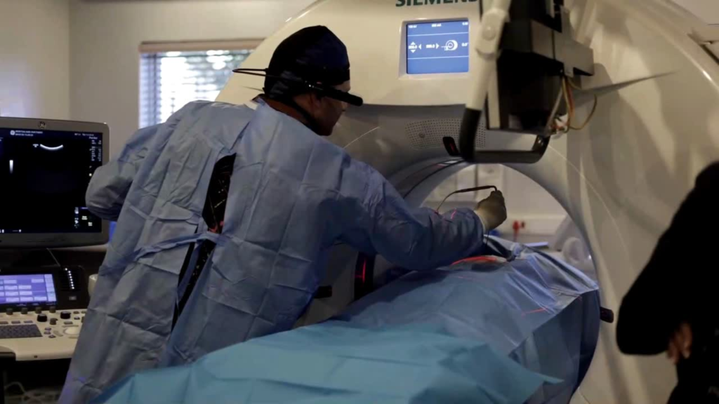
In this video, Dr Frikkie Buitendag performs a recent cryoablation procedure. It shows how the needles are guided into place and how the ice ball forms during treatment.

One message to remember

Asked for the one thing listeners should remember, Dr Buitendag said:

“Keep all your options in mind and ask your referring doctor or oncologist what they are. Reading about these things on the internet can cause a lot of anxiety, so get a professional opinion on what is best for you. Surgery and the smaller procedures both have advantages and disadvantages. It isn’t always suitable, but it is an option worth considering. And it shouldn’t be decided by the radiologist alone. Everyone should work together in a multidisciplinary team, so that we’re all on the same page and doing what is best for the patient.”

Listen to the full interview

The full interview (in Afrikaans) is available from RSG’s Gesondheid programme. Listen on RSG

If you have been diagnosed with a small kidney, liver or lung lesion, ask your urologist or oncologist whether ablation could be an option for you.

Referring doctors can contact Morton & Partners’ interventional radiology team at cathlab@morton.co.za.